
A method of nerve regeneration after spinal cord injury has been developed
An international team of researchers has developed a three-step method for regenerating severed nerve fibers after spinal cord injury. Tests on rodents have yielded very promising results, and if similar results can be obtained in humans, this could lead to an effective therapy for treating spinal injuries.
Spinal cord injuries lead to a situation in which theorej severed trichomesoNerve spines do not regenerate in the central nervous system. As a result, electrical signals from the mozg pertaining to body movement no longer reach the muscles, leading to complete and permanent paralysis. To counteract this, the researchers developed a therapy method thatora causes the re-growth of axonow. The new treatment has yielded extremely promising results. At least in tests on mice.
A new three-step treatment has been developed by scientists of theow from the University of California at Los Angeles (UCLA), Ecole polytechnique fédérale de Lausanne (EPFL) and from Harvard University. The therapy causes axons – tiny fibers of theokna, whichore combine comork nerves and enable them to communicate, regenerate after spinal cord injury. In rodent tests, the axons not only broke through the scars, but row also could carry signals through the damaged tissue.
If researchers are able to get similar results in human studies, the findings could lead to regeneration of axon connectionsoin the u osob after spinal cord injury, potentially restoring all their functions. The results of the experiments were published on „Nature”.
– The idea was to combine three rodifferent therapies and see if this combination could stimulate regeneration of severed axonsow for re-growth through spinal cord injury scars – said the head of theown author of the publication Michael Sofroniew, professor of neurobiology at UCLA. – Previous studies had tested each of the three therapies separately, but never together, and this proved crucial – added.
Spinal cord rupture leads to axon damageoin and preventing mozgowi sending a signaloin to the neuronow below the site of injury. This leads to paralysis and loss of other neurological functions. The scientific approachow from UCLA may be the first step to solving this problem.
This three-step method was developed to restore the conditions of theoin the underlying growth of the trichomeonerve trunks during fetal development, but it has been shown to help regenerate severed fibersonerve trunks following complete spinal cord injury.
– Our goal was to reproduce in adults the conditions ofoin areas conducive to hair growthonerve cells during development. We have understood the combinations of biological mechanismow, whichore are necessary to allow regrowth of the haironerve trunks – admitted Grégoire Courtine of EPFL, coopublication author.
To explain the method developed by the researchers, Courtine used the analogy of trees. If the trawlonerve spines would be trees, woat the time the terminaloaxonow could be viewed as branches of a tree. If the headown tree branches will be cut off, small branches can spontaneously grow along the remaining tree trunk, but the cut branches never grow back. The same goes for the neuronow in adults: new branches of severed axonsow can sprout and form connections above the injury, but the severed portion of the axon does not grow back. However, a new method changes this and makes it possible to regenerate whole axonsow.
Decades of research have shown that to the growth of human włonerve cells require three things. These are genetic instructions to activate axon growthow, a molecular pathway that determines the pathway along which theorej axons to regrow and the right nutrient for the axonoin stimulating them to regenerate. All these conditions are met when a person develops in the womb. After birth, these processes disappear, but the genes controlling the process are still present.
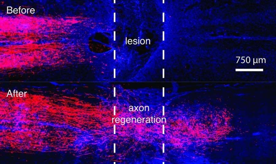
The researchers succeeded in awakening the corresponding geneow and resumption of this process. Not only have the axons grown through scar tissue, but many have formed new connections to neurons and have shown signs of electrical activity.
– When we stimulated the animal’s spinal cord with a low electric current over the site of injury, the regenerated axons showed about 20 percent of the. normal electrical activity below the lesion,” said Sofroniew.
In tests on mice, axons grew back at a rate of a few millimetersoin for a month, but as the scientists pointed out, the regenerated axons are able to communicate, but this regained connectivity is not enough to restore theocic walking. Rehabilitation with electrical stimulation is required to integrate and fine-tune the new axonsow to restoreorestore lost functions.
– We expected these regenerated axons to behave like axons during development – not immediately support coordinated function. Just as a newborn baby must learn to walk, axons thatore regrowth after injury will require training – Sofroniev explained.
This is certainly quite an achievement, but it is still premature to speculate on applications in humans – researchers emphasize. More research is needed for this method to be tested on humans.